Congrats to Ellen and Sara on the recent publication!

Multiplexed silicon photonic sensor arrays enable facile characterization of coagulation protein binding to Nanodiscs with variable lipid content

ABSTRACT

Interactions of soluble proteins with the cell membrane are critical within the blood coagulation cascade. Of particular interest are the interactions of γ-carboxyglutamic acid-rich (GLA) domain-containing clotting proteins with lipids. Variability among conventional analytical methods presents challenges for comparing clotting protein-lipid interactions. Most previous studies have investigated only a single clotting protein and lipid composition and have yielded widely different binding constants. Herein, we demonstrate that a combination of lipid bilayer Nanodiscs and a multiplexed silicon photonic analysis technology enables high-throughput probing of many protein-lipid interactions among blood-clotting proteins. This approach allowed direct comparison of the binding constants of prothrombin, factor X, activated factor VII, and activated protein C to seven different binary lipid compositions. In a single experiment, the binding constants of one protein interacting with all lipid compositions were simultaneously determined. A simple surface regeneration then facilitated similar binding measurements for three other coagulation proteins. As expected, our results indicated that all proteins exhibit tighter binding (lower Kd) as the proportion of anionic lipid increases. Interestingly, at high proportions of phosphatidylserine, the Kd values of all four proteins began to converge. We also found that although koff values for all four proteins followed trends similar to those observed for the Kd values, the variation among the proteins was much lower, indicating that much of the variation came from the kinetic binding (kon) of the proteins. These findings indicate that the combination of silicon photonic microring resonator arrays and Nanodiscs enables rapid interrogation of biomolecular binding interactions at model cell membrane interfaces.

Click here to read the manuscript.

Congrats James, Colleen and Josh on the recent publication!


Microfluidic Platform for Efficient Nanodisc Assembly, Membrane Protein Incorporation, and Purification

ABSTRACT

The characterization of integral membrane proteins presents numerous analytical challenges on account of their poor activity under non-native conditions, limited solubility in aqueous solutions, and low expression in most cell culture systems. Nanodiscs are synthetic model membrane constructs that offer many advantages for studying membrane protein function by offering a native-like phospholipid bilayer environment. The successful incorporation of membrane proteins within Nanodiscs requires experimental optimization of conditions. Standard protocols for Nanodisc formation can require large amounts of time and input material, limiting the facile screening of formation conditions. Capitalizing on the miniaturization and efficient mass transport inherent to microfluidics, we have developed a microfluidic platform for efficient Nanodisc assembly and purification, and demonstrated the ability to incorporate functional membrane proteins into the resulting Nanodiscs. In addition to working with reduced sample volumes, this platform simplifies membrane protein incorporation from a multi-stage protocol requiring several hours or days into a single platform that outputs purified Nanodiscs in less than one hour. To demonstrate the utility of this platform, we incorporated Cytochrome P450 into Nanodiscs of variable size and lipid composition, and present spectroscopic evidence for the functional active site of the membrane protein. This platform is a promising new tool for membrane protein biology and biochemistry that enables tremendous versatility for optimizing the incorporation of membrane proteins using microfluidic gradients to screen across diverse formation conditions.

Click here to read the manuscript.

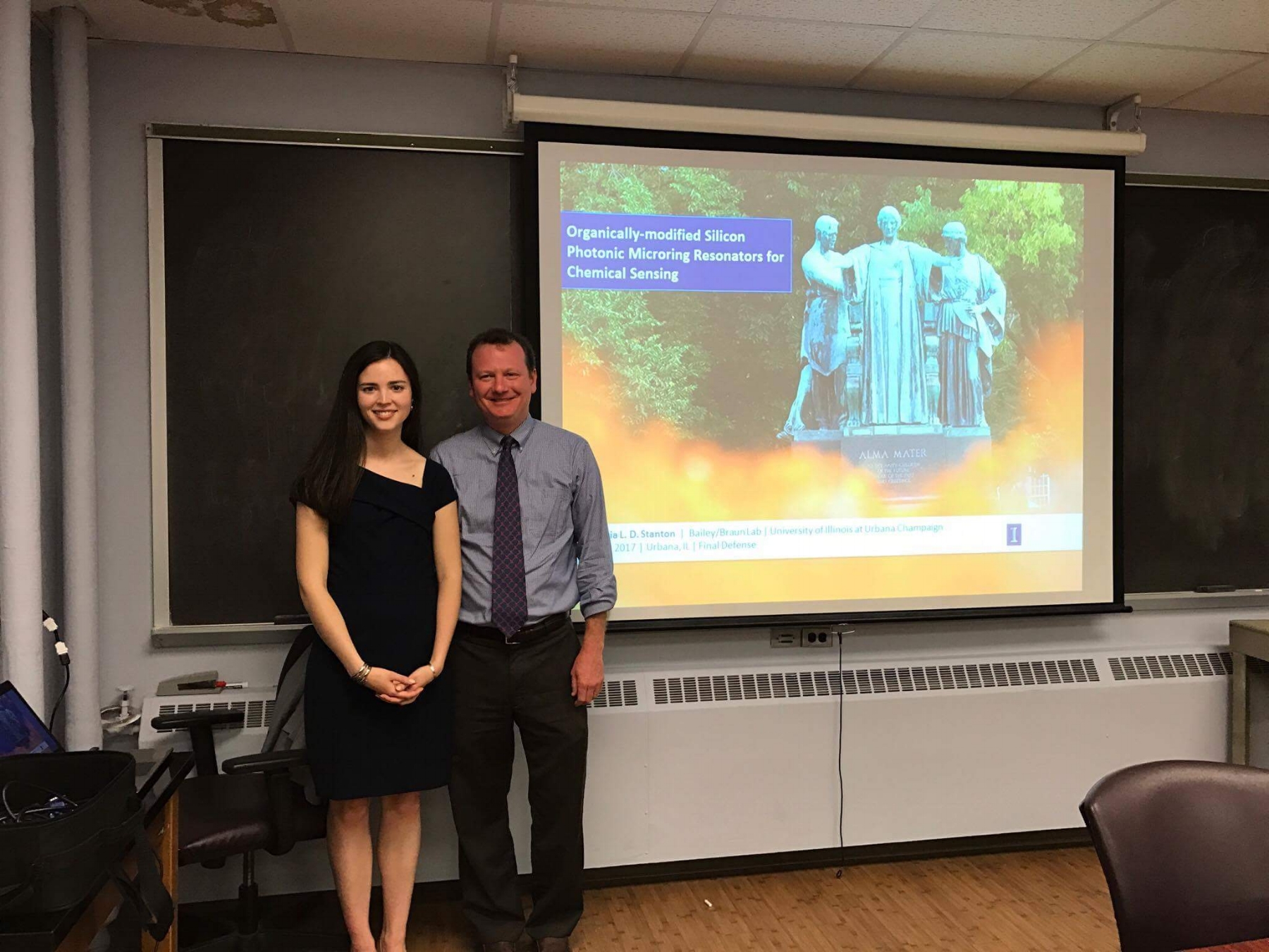
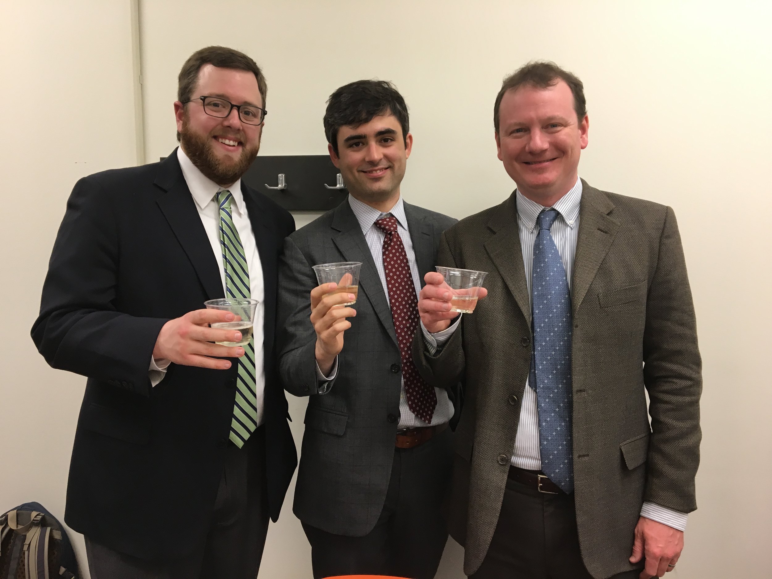
Congrats to Dr. Graybill and Dr. Wade!

They each successfully defended their theses, though the cake may have suggested a different celebration. Congratulations to Dr. Graybill for successfully defending his thesis "Improving Multiplexed RNA Detection Assays By Interfacing Enzymatic Amplification Strategies with Silicon Photonic Microring Resonators.” Congratulations to Dr. Wade, as well. He defended his thesis "Expanding the Toolbox for Personalized Medicine with Silicon Photonic Microring Resonators and Microfluidic Technologies."

IMG_3499.JPG

Congrats to Steve on his recent publication!

The K-Channel: A Multi-Functional Architecture for Dynamically Re-Configurable Sample Processing in Droplet Microfluidics

ABSTRACT

By rapidly creating libraries of thousands of unique, miniaturized reactors, droplet microfluidics provides a powerful method for automating high-throughput chemical analysis. In order to engineer in-droplet assays, microfluidic devices must add reagents into droplets, remove fluid from droplets, and perform other necessary operations, each typically provided by a unique, specialized geometry. Unfortunately, modifying device performance or changing operations usually requires re-engineering the device among these specialized geometries, a time-consuming and costly process when optimizing in-droplet assays. To address this challenge in implementing droplet chemistry, we have developed the “K-channel,” which couples a cross-channel flow to the segmented droplet flow to enable a range of operations on passing droplets. K-channels perform reagent injection (0-100% of droplet volume), fluid extraction (0-50% of droplet volume), and droplet splitting (1:1-1:5 daughter droplet ratio). Instead of modifying device dimensions or channel configuration, adjusting external conditions, such as applied pressure and electric field, selects the Kchannel process and tunes its magnitude. Finally, interfacing a device-embedded magnet allows selective capture of 96% of dropletencapsulated superparamagnetic beads during 1:1 droplet splitting events at ~400 Hz. Addition of a second K-channel for injection (after the droplet splitting K-channel) enables integrated washing of magnetic beads within rapidly-moving droplets. Ultimately, the K-channel provides an exciting opportunity to perform many useful droplet operations across a range of magnitudes without requiring architectural modifications. Therefore, we envision the K-channel as a versatile, easy to use microfluidic component enabling diverse, in-droplet (bio)chemical manipulations.

Click here to read the manuscript.

Congrats to Alex on Her Recent Publication

Polymer Brush-Modified Microring Resonators for Partition-Enhanced Small Molecule Chemical Detection, has been accepted for publication in Wiley-VCH's newest journal, ChemistrySelect. This article is the result of a collaboration with Paul Braun's Materials Science and Engineering group at the University of Illinois at Urbana-Champaign and introduces the idea of organically modifying our ring resonators for specific and selective detection.

Congrats to Dan on his most recent paper published in Langmuir!

Dan's most recent paper published in Langmuir. This is an extension of his previous work published last year, which demonstrated nanoporous gold (NPG) as a material with selective and tunable chemical transport properties.

Electrolyte Gradient-Based Modulation of Molecular Transport through Nanoporous Gold Membranes

Abstract

Nanopores, and nanoporous materials in general, are interesting for applications in chemical and biomolecular transport as pore sizes are on the same scale as the dimension of many (bio)chemical species. Many studies have focused on either single pores or small arrays of cylindrical pores, which are convenient in terms of their amenability toward computational modeling of transport phenomenon. However, the limited overall porosity may inhibit transport flux as well as the eventual implementation of these materials as active separation elements. Inspired by its relatively high porosity, we have explored nanoporous gold (NPG) as a membrane across which small molecular species can be transported. NPG offers a random, bicontinuous pore geometry, while also being inherently conductive and readily amenable to surface modification—attributes that may be enabling in the pursuit of size- and charge-based approaches to molecular separations. NPG was fabricated via a free-corrosion process whereby immersion of Au-containing alloys in concentrated nitric acid preferentially dissolves the less noble metals (e.g., Ni, Cu). Average pore diameters of 50 ± 20 nm were obtained as verified under scanning electron microscopy. NPG membranes were sandwiched between two reservoirs, and the selective transport of chemical species across the membrane in the presence of an ionic strength gradient was investigated. The flux of small molecules were monitored by UV–vis absorption spectrometry and found to be dependent upon the direction and magnitude of the ionic strength gradient. Moreover, transport trends underscored the effects of surface charge in a confined environment, considering that the pore diameters were on the same scale as the electrical double layer experienced by molecules transiting the membrane. Under such conditions, the transport of anions and cations through NPG was found to depend on an induced electric field as well as ion advection. Further electrical and surface chemical modulations of transport are expected to engender increased membrane functionality.